Centre Nacional d'Anàlisi Genòmica
Centro Nacional de Análisis Genómico
Centro Nacional de Análisis Genómico
BCN, 18 Feb, 2021.- Researchers at the CNAG-CRG and IDIBAPS-Hospital Clínic in Barcelona have led a study that charts the first three-dimensional genomic map of of healthy and cancerous lymphocytes. The team have developed a new way to map the three dimensional structure of the human genome, revealing hundreds of alterations in leukaemias and lymphomas.
The study was coordinated by ICREA researchers Iñaki Martín-Subero from IDIBAPS and Marc A. Marti-Renom with dual affiliation between the Centro Nacional de Análisis Genómico (CNAG-CRG) and the Centre for Genomic Regulation (CRG). The first authors of the study, published in the journal Nature Communications, are Roser Vilarrasa-Blasi and Paula Soler-Vila, postgraduate students from both laboratories.
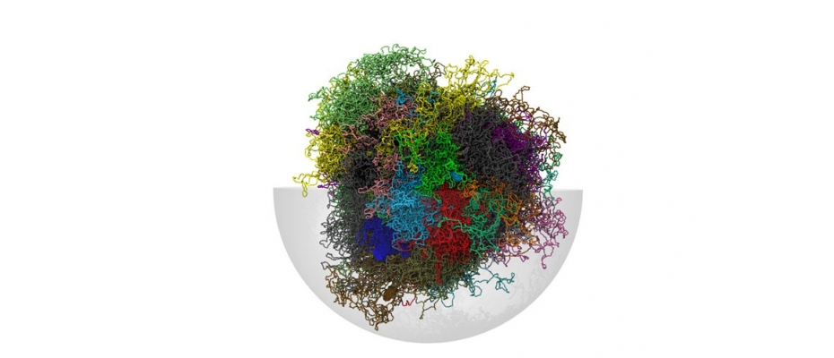
If we stretched the human genome in each of our cells it would measure two metres. All this DNA is packaged within the cell nucleus, forming a complex three-dimensional network. This three-dimensional structure plays an important role in the function of a cell, determining which genes are expressed and which are silenced.
Using advanced molecular biology and bioinformatics methods, researchers have described the first three dimensional maps of the lymphocyte maturation process, as well as for two types of lymphoid cancer, chronic lymphocytic leukemia and mantle cell lymphoma.
The ambitious project came together though the collaboration between Martin-Subero’s research group, an expert in epigenetics of haematological tumours, and Marti-Renom’s group, expert in structural genomics. According to Roser Vilarrasa-Blasi and Paula Soler-Vila, “neither of the two research groups had enough experience to carry out this study individually. We worked side by side for several years to draw and interpret 3D maps of healthy and tumorous lymphocytes. This study is a great example of the power of collaboration and communication between experts from different disciplines”.
Previous studies had identified that the genome within the cell nucleus separates into two spaces, an active compartment and an inactive compartment, and three-dimensional maps were defined using this classification. According to Iñaki Martin-Subero, “when analysing the data of the 3D interactions of the genome and correlating them with other epigenetic marks, we realized that it is more appropriate to classify the genome into three spatial components; the active, the inactive and a new highly dynamic intermediate compartment that represents the bridge that communicates between spaces”.
Researchers found unexpected results when studying the genomic architecture changes in the lymphocyte maturation process. “We have observed that approximately one third of the 3D interactions of the genome change during the physiological process of lymphocyte maturation, which indicates that the 3D map of the genome is very dynamic”. In leukaemia and lymphomas, Marti-Renom continues, “we focus on the fraction of the genome that does not change in normal lymphocytes in order to detect changes specific to leukemias and lymphomas. We were able to observe that large blocks of DNA change their 3D structure, and these blocks contain important genes for the development of tumors”, says Marc A. Marti-Renom.
According to the researchers, knowing the three-dimensional map of the leukaemia and lymphoma genome is one more step towards a better understanding of the molecular basis of cancer, but there is still a long way to go before we can talk about clinical applications. “It is important to emphasize the importance of basic cancer research in order to later translate findings into clinical improvements. In this case, alterations in the 3D structure of the genome constitute a new axis of vulnerability for tumour cells and are a promising therapeutic target", says Iñaki Martin-Subero.
Work of reference